Back to News

Incidence of Spontaneous and Urethane-Induced Tumors in a 26-Week Carcinogenicity Study in Tg.rasH2 Mice Sourced From CLEA, Japan

November 04, 2025

Abstract
The Tg.rasH2 mouse is a validated bioassay system for evaluation of carcinogenic potential, and it is confirmed to be sensitive to both genotoxic and nongenotoxic carcinogens. This is also one of the models specified in ICH S1B Guidance, enabling 6-month carcinogenicity studies as an alternative to traditional 2-year bioassays. We conducted a 26-week carcinogenicity study at our test facility on Tg.rasH2 mice sourced from CLEA Japan Inc., in a process to generate historical control database. Although historical control data have been published for these mice sourced from Taconic Biosciences Inc., USA, there is a dearth of published literature citing spontaneous incidence of neoplastic findings in Tg.rasH2 mice sourced from CLEA Japan. We have therefore presented the spontaneous tumor incidence in our study and compared it with the previously published tumor incidence for Tg.rasH2 mice sourced from Taconic, USA. The comparison reveals a similarity of tumor incidence between the mice from these two sources.
Introduction
The Tg.rasH2 mouse model has now established its validity for evaluation of carcinogenic potential in 26-week bioassays. These mice have been confirmed to be sensitive to both genotoxic and nongenotoxic carcinogens.1.The Tg.rasH2 model has been proven to predict neoplastic findings relevant to human cancer risk assessment, produces fewer non-biologically significant neoplastic outcomes, and is thus preferable to a 2-year rodent study.2 It is one of the transgenic mouse models discussed at length in the ICH Guidance for industry in its S1B(R1) addendum to S1B testing for carcinogenicity of pharmaceuticals, enabling 6-month carcinogenicity studies as an alternative to traditional 2-year bioassays.3
The rasH2 mouse was created by Dr Motoya Katsuki and others under the supervision of Director Tatsuji Nomura of the Central Institute for Experimental Animals (CIEA, a Public Interest Foundation; it was renamed in April 2024 as CIEM—Central Institute for Experimental Medicine and Life Science). In 1992, the institute began practical application of carcinogenicity evaluation tests, and the high reproducibility and stability of the carcinogenicity evaluation of the rasH2 mouse was demonstrated by an international collaborative research project by the International Life Sciences Institute (ILSI)/Health and Environmental Sciences Institute (HESI) involving 50 industrial, governmental, and academic facilities from Japan, the United States, and Europe over a 4-year period from 1997 to 2000. It was concluded that the CB6F1-TgrasH2 mouse appeared to be a promising model for short-term carcinogenicity studies. CLEA (Central Laboratory for Experimental Animals) began full-scale production and supply in 2001.4 Tg.rasH2 is a genetically modified mouse that has been introduced with the human-derived proto-oncogene c-Ha-ras. Spontaneous tumor development is minimal up to 34 weeks of age. Test substances are administered starting at around 8 weeks of age, and tumor development is evaluated over a 26-week period, making it possible to determine whether they are carcinogenic.4 These spontaneous tumors include bronchiolo-alveolar adenoma/adenocarcinoma in the lung, hemangiosarcoma in the spleen and various other tissues, adenoma and adenocarcinoma of the Harderian gland, rhabdomyosarcoma in the subcutaneous tissue, and thymoma. Rapid induction of more malignant tumors was observed after treatment with various carcinogens.5 The transgenic rasH2 mice were found to be generally much more susceptible to both mutagenic and non-mutagenic carcinogens than were non-Tg mice, pointing to advantageous application for detection of carcinogenic potential.6
Worldwide, the breeding and supply of Tg.rasH2 mice are restricted to two sources: Taconic Biosciences Inc. and CLEA Japan Inc. The Tg.rasH2 mice derived from Taconic are mostly used for studies conducted in the US and Europe. Published historical control data are available for almost one hundred 26-week carcinogenicity studies conducted using the Tg.rasH2 mice supplied by Taconic Biosciences Inc.7 The CLEA bred Tg.rasH2 mice are used for conducting studies in Asia and other countries. However, since there are no similar publications of historical control data available for these mice, it is difficult to estimate the number of studies that have been conducted using CLEA-derived Tg.rasH2 mice.
Two positive control materials have been used in the 26-week Tg.rasH2 studies to prove the validity of the assays. N-methyl-N-nitrosourea (MNU) was used as a positive control material by various laboratories, where the animals were kept on the experiment for 14 weeks after the start of administration.5 The most common neoplastic lesions induced by MNU are forestomach neoplasms histologically observed as squamous cell papilloma or carcinoma. Other common MNU-induced neoplastic lesions are malignant lymphoma mainly composed of thymic lymphoma in the hematopoietic system, bronchiolo-alveolar adenoma, or carcinoma in the lungs, and squamous cell papilloma, carcinoma, and keratoacanthoma on the skin.8 It is worthwhile to note that the papilloma and lymphomas are not the spontaneous tumors of Tg.rasH2.
Tg.rasH2 mice treated with the positive control urethane produce lung tumors and splenic hemangiosarcomas, which are also the spontaneous tumors of these mice. These tumors are more relevant to human beings and involve both epithelial and mesenchymal cell lines. As a part of process improvement and with adherence to the 3Rs of animal research, the scientists at BioReliance not only reduced the experimentation period from 26 weeks to 12 weeks for the positive control groups but also reduced the number of animals to 10 in each sex.9
Urethane, when dosed at 1000 mg/kg bwt, has been observed to induce untoward clinical signs of toxicity/exaggerated pharmacological effects in the treated mice.11,12 A study by Paranjpe et al (2021)10 reported testing of a lower dose of 500 mg/kg bwt of urethane on days 1, 3, and 5, in Tg.rasH2 mice sourced from Taconic, and demonstrated reduction in the toxicity without affecting the incidence of tumors. The urethane-treated positive control Tg.rasH2 mice are maintained on the study for 10–12 weeks, and upon termination only the lungs and spleen are evaluated microscopically.10 Because of these obvious differences, use of urethane as a positive control substance is considered better in Tg.rasH2 studies than MNU.
Material and Methods
Animals
A total of 120 number of CByB6F1-Tg(HRAS)2Jic mice, 60 males and 60 females, were purchased from CLEA Japan Inc. (1-2-7 Higashiyama, Meguro-Ku Tokyo, Japan). The mice were 4 to 5 weeks old when received at the test facility of Intox Pvt Ltd, in Pune, India, and underwent a one week of quarantine before they were allocated for the present study. The mice were acclimated to the experimental room for a period of at least one week prior to the first day of treatment.
Housing Feeding and Environmental Conditions
The mice were housed in individually ventilated polycarbonate cages with gamma-irradiated Aspen Shavings Wooden Chips bedding in environmentally controlled rooms. Animals were verified to be healthy prior to being placed on the study. All animals had ad libitum access to gamma-irradiated “Altromin” brand pelleted rodent feed manufactured by M/s Altromin Spezialfutter GmbH & Co. KG, Germany. Potable water that was passed through “Aquaguard” water filter and autoclaved was provided ad libitum in sterilized water bottles with stainless steel sipper tubes. The diet, drinking water, and the bedding were tested and certified to be free from undesired levels of environmental contaminants.
Experimental Design
Of a total of 120 number of Tg.rasH2 mice, 100 mice (50 per sex) served as the untreated control group and were used for assessment of the incidence of spontaneous tumors. The remaining 20 mice (10 per sex) were assigned to the positive control group. Each mouse was assigned a unique identification (ID) number. The mice were 6–7 week old at their first dose of urethane. The average weights of the male and female mice from the urethane treated group were 23.28 g and 18.87 g, respectively.
The mice from the positive control group were injected with 500 mg/kg bwt of urethane (alkylating genotoxic carcinogen) intraperitoneally on days 1, 3, and 5. The urethane (Sigma-Aldrich Chemical Pvt Ltd, Lot No. WXBD1167 V) was dissolved in saline (sodium chloride −0.9% w/v) at a concentration of 50 mg/mL, and the intra-peritoneal injections to the mice were made using insulin syringe at a dosage volume of 10 mL/kg bwt.
In-Life Observations
The mice were observed twice daily for survival and for any abnormal signs, at least 6 h apart. Their food consumption was recorded, and daily food intake was calculated. Body weights of the mice were initially recorded weekly up to 13 weeks and thereafter biweekly until the terminal necropsy. All mice were fasted for 3–4 h prior to the scheduled necropsy.
Study Termination and Pathology

The 20 mice allocated to the positive control group were sacrificed at the end of 16 weeks of the experimental period, while rest of the 100 mice from the untreated control groups were sacrificed at the end of the 26-week observation period. The mice were euthanized by carbon dioxide asphyxiation followed by exsanguination. A complete necropsy was performed on any mice that were found dead or sacrificed moribund during the study, and on those that were sacrificed at study termination. Two mice were found dead, one male mouse on day 118 and one female mouse on day 141. All tissues listed in Table 1 were collected, fixed in 10% neutral buffered formalin, embedded in paraffin, sectioned, stained with hematoxylin and eosin, and evaluated microscopically. Testes and eyes with optic nerves were preserved in modified Davidson’s fixative. For the urethane-treated positive control animals, only the expected target organs (lungs and spleen) were evaluated microscopically. Microscopic images were captured by using the OLYMPUS DP74 camera system installed on OLYMPUS BX43 microscope.

Table 1. List of Tissues Evaluated Microscopically in Untreated Control Tg.rasH2 Mice.
Adrenal Glands Kidneys With Ureter Pituitary Gland Testes
Aorta Large intestine Prostate gland Thymus
Bone Lacrimal gland Salivary glands Thyroid glands
Brain Liver Sciatic nerve Tongue
Clitoral gland Lungs Seminal vesicles Trachea
Epididymides Lymph nodes (mandibular and mesenteric) Skeletal muscle Urinary bladder
Esophagus Mammary gland (female) Skin Uterus with cervix
Femur with joint Nasal cavity Small intestine Vagina
Eyes with optic nerve Ovaries with oviduct Spinal cord Zymbal gland
Gall bladder Pancreas Spleen Gross lesions
Harderian gland Parathyroid glands Sternum with bone marrow
Heart Preputial gland Stomach
Pathology peer review was conducted to verify and endorse the accuracy and consistency of diagnostic terminology used in the study.
Comparison With Published Data
The data on spontaneous incidence was then compared with similar data published by Paranjpe et al7 wherein Tg.rasH2 mice sourced from Taconic, USA, were tested in the 26-week carcinogenicity studies conducted between 2004 and 2012 (710 mice/sex) and between 2014 and 2018 (905 male and 850 female mice). Additionally, the data was also compared for studies conducted between 2014 and 2023 (46 male and 47 female studies) by the Labcorp Early Development Laboratories, Madison, WI site. These mice were also sourced from Taconic. Similarly, the incidence of urethane-induced neoplastic findings was compared with two other publications (Paranjpe et al10; Ambroso et al11).
Regulatory Compliances
The study was performed in full compliance with the OECD Principles of Good Laboratory Practice (OECD, 1998) and followed the Study Plan (Protocol) and the standard operating procedures in force at the AAALAC-accredited Test Facility of Intox Pvt Ltd. The study protocol was approved by the Institutional Animal Ethics Committee (Form B-B08), while the study was performed under the conditions recommended by the Committee for Control and Supervision of Experiments on Animals (CCSEA) of the Government of India (its “Breeding of and Experiments on Animals (Control and Supervision) Amendment Rules,” 2006) and by the US publication by National Research Council, Institute of Laboratory Animal Resources, 2011, “Guide for the Care and Use of Laboratory Animals,” National Academy Press, Washington, DC.
Results
Spontaneous Neoplastic Findings
Lung Adenomas and Adenocarcinomas
Bronchiolo-alveolar adenomas of the lungs were the most common spontaneous tumors in both sexes of mice, with an incidence of 10% in males and 8% in females. Distinct, tan-colored raised nodules (0.2 × 0.5 cm) on the lung surface were predominantly adenomas, while bronchiolo-alveolar adenocarcinomas were larger masses occupying part of the lobule or multiple lobules. Microscopically, the adenomas were characterized by neoplastic cells which were cuboidal to columnar and formed papillary projections or compact acinar structures. The cells were well differentiated, had uniform round to oval nuclei, and abundant cytoplasm with distinct cellular outlines (Figure 1). Similarly, bronchiolo-alveolar carcinomas were characterized by multiple layers of proliferating epithelial cells seen forming solid clusters or sheets of cells. The cells had indistinct cellular outlines and marked cellular atypia characterized by pleomorphism. The nuclei were round to oval with a moderate degree of mitosis. Invasion of surrounding parenchyma was also seen (Figure 2).
Figure 1. Representative microphotograph of bronchiolo-alveolar single well-circumscribed adenoma (black arrow) with surrounding normal tissue (N) at 40X.
Figure 2. Representative microphotograph of bronchiolo-alveolar adenocarcinoma with indistinct cellular outlines and mitosis (black arrows) at 200X.
The incidence of bronchiolo-alveolar adenocarcinomas was 4% and 0% in male and female mice, respectively. The combined incidence of all primary pulmonary tumors (adenomas and adenocarcinomas) was 14% in males and 8% in females.
Splenic Hemangiosarcoma
Splenic hemangiosarcoma was the second most common spontaneous neoplastic lesion in the control Tg.rasH2 mice with incidence of 2% in males and 4% in females. Grossly, the splenic hemangiosarcomas were observed as nodules, 3 to 5 mm in diameter, either red or tan in color, or as depressed foci of similar color and dimensions. Microscopically, malignant endothelial cells in hemangiosarcomas often lined the vascular spaces containing blood. Endothelial cells had polygonal to spindeloid hyperchromatic nuclei with scanty cytoplasm. The cellular outlines were distinct. No metastasis of these tumors in the peritoneal cavity was observed (Figure 3). For comparative histological evaluation, Figure 4 illustrates the normal spleen architecture.
Figure 3. Representative microphotograph of splenic hemangiosarcoma replacing normal splenic tissue with vascular spaces containing blood at 100X.
Figure 4. Representative microphotograph of normal spleen at 100X.

Spontaneous Hemangiosarcomas in Organs Other Than Spleen

There were single instances of such hemangiosarcomas observed each in males (testes; incidence 2%) and in females (uterus; incidence 2%) (Figure 5). No hemangiomas were observed. For comparative histological evaluation, Figure 6 illustrates the normal uterine architecture.
Figure 5. Representative microphotograph of uterine hemangiosarcoma with vascular proliferation invading the endometrium at 100X.
Figure 6. Representative microphotograph of normal uterus at 100X.

Other Spontaneous Tumors

There were a few non-pulmonary and non-vascular spontaneous neoplastic findings such as squamous cell carcinoma in the submandibular salivary gland, keratoacanthoma in skin of the head and rhabdomyoma in skeletal muscles, adenoma (Figure 7) and adenocarcinoma of Harderian glands (Figure 8), and thymoma in thymus (Figure 9). For comparative histological evaluation, Figure 10 illustrates the normal thymic architecture.
Figure 7. Representative microphotograph of well-demarcated Harderian gland adenoma (black arrow) slightly compressing the surrounding normal tissue (N) at 40X.
Figure 8. Representative microphotograph of Harderian gland adenocarcinoma with pleomorphic cells (black arrow) and cytoplasmic vacuolation (yellow arrow) at 100X.
Figure 9. Representative microphotograph of thymoma with lymphocytes admixed with spindle-shaped cells (black arrow) at 100X.
Figure 10. Representative microphotograph of normal thymus at 100X.

Body Weights

Body weights of the mice were initially recorded weekly up to 13 weeks and thereafter biweekly until the terminal necropsy. The average body weights of Tg.rasH2 diet control male and female mice at the start of the study were 21.2 g and 16.9 g, respectively. Individual body weights for the diet control group of each sex were within ±20 % of the mean at the start of the study. The average body weights of Tg.rasH2 diet control male and female mice at the termination of the 6-month studies were 26.7 and 20.8 g, respectively.

Incidence of Neoplastic Findings in Urethane-Treated (Positive Control) Tg.rasH2 Mice

The mice treated with urethane at 500 mg/kg body weight revealed a few clinical signs like hunched posture, lethargy, and labored breathing. There was also a transient reduction in their body weight gain during the first week of the study.

The incidence of neoplastic findings in the mice in this group has been presented in Table 2 and in the ensuing text.

Table 2. Incidence of Neoplastic Findings in Positive Control Group Mice.
Sex of Tg.rasH2 mice Male Female
Number of mice/group 16 16
Duration of weeks on study 16 16
Organ/tissue Numbers % Numbers %
Lungs
 Bronchiolo-alveolar adenoma, multiple 10 100 10 100
 Bronchiolo-alveolar adenocarcinoma 3 30 4 40
 Hemangiosarcoma 1 10 2 20
Spleen
 Hemangiosarcoma 10 100 10 100

Tumors in Lungs

The gross examination revealed multiple (1–2) nodules in lungs of all the 20 mice (100% incidence) and the same were microscopically confirmed (1–2 tumors per lung) to be bronchiolo-alveolar adenomas/adenocarcinomas. Also, the lungs in 3/20 mice revealed hemangiosarcomas co-existing with bronchiolo-alveolar adenomas/adenocarcinomas.

Tumors in the Spleen

At necropsy, the spleens revealed nodules (1–2) in 18/20 mice. Upon microscopic examination, these were confirmed to be hemangiosarcomas and were also evidenced microscopically in additional two mice, thereby reporting their incidence in all the 20/20 mice (incidence of 100%).

Discussion

The acceptance of alternate mouse transgenic models such as Tg. rasH2 and p53+/− mice for short-term (ie, 6-month) cancer biohazard testing of pharmaceuticals by the FDA and ICH has been an important milestone from drug development and human risk assessment perspectives.11 Some of the factors contributing to limited use of these models by the biopharmaceutical industry had been concerns regarding lack of historical data, application of consistent diagnostic criteria, use of non-standardized terminologies in published data, and lack of comfort using a novel model.13
Caution should be exercised in using historical control data published from other laboratories, as stated by the US Food and Drug Administration.14 Since interlaboratory differences in spontaneous tumor incidence have been previously reported,15 it would be appropriate to use a database generated in-house and, if possible, do a retrospective analysis with the published data. Therefore, the present 6-month carcinogenicity study was conducted in the test facility of Intox to understand the incidence of the spontaneous and urethane (positive control)-induced neoplastic alterations in rasH2 mice and to generate laboratory-specific historical database.
The data on spontaneous tumor incidence in the present study was compared with similar data published by Paranjpe et al7 in their two publications which present historical control data for spontaneous tumors in transgenic rasH2 mice sourced from Taconic, USA. Our data was also compared with studies conducted at the Labcorp Early Development Laboratories site in Madison, WI.16 These mice were likewise sourced from Taconic. The comparison made in the present manuscript, however, must be seen considering the large differences in the number of mice on which these two published databases rest on, and only the 50 mice/sex evaluated in the present study at Intox. However, for the positive control data, the comparison of incidence of urethane-induced neoplastic findings in 20 mice in the present study was made with the incidence described in the earlier publication of Paranjpe et al.10

Comparative Incidence of Common Spontaneous Neoplastic Findings

Tables 3 and 4 present the comparative incidence of common spontaneous neoplastic findings at Intox, which in general are seen to be comparable to the published databases. It may be noted that the comparison could not be made using any statistical tools in absence of data from individual studies. Certain important trends in tumor incidence, such as the most common spontaneous tumors, tissues/organs with higher and lower incidence of tumors, and the types of tumors, were found to be similar in the Intox’s study and the two published databases. This was despite the smaller number of mice tested in the study of Intox.

Table 3. Comparative Incidence of Common Spontaneous Neoplastic Findings in Male Mice Sourced From CLEA and TACONIC.
Data source Current study (2023) Paranjpe et al7 Labcorp16
Source of mice CLEA Taconic Taconic Taconic
Period 2023 2004–2012 2013–2018 2014–2023
Sex Male Male Male Male
No. of mice/sex 50 710 905 1525
Duration of weeks on study 26 26 26 26
Tissue/organ and finding Incidence of spontaneous neoplastic findings %
Number of mice % Number of mice % Number of mice % Number of mice
Lungs
 Bronchiolo-alveolar adenoma 5 10.00 83 11.69 77 8.51 96 6.3
 Bronchiolo-alveolar adenocarcinoma 2 4.00 4 0.56 20 2.21 24 1.6
 Lung tumors combined 7 14.00 87 12.25 97 10.72 120 7.9
Spleen
 Hemangiosarcoma 1 2.00 26 3.66 35 3.87 58 3.8
Testes
 Hemangiosarcoma 1 2.00 5 0.70 2 0.22 0 0
Harderian glands
 Adenoma 0 0.00 10 1.41 12 1.33 11 0.7
 Adenocarcinoma 1 2.00 1 0.14 13 1.44 5 0.3
Thymus
 Thymoma, benign 0 0.00 1 0.14 0 0.00 10a 0.7
Salivary glands
 Submandibular, squamous cell carcinoma 0 0.00 1 0.14 0 0.00 0 0.00
Skin (head)
 Keratoacanthoma, benign 1 2.00 0 0.00 0 0.00 0 0.00
Skeletal muscles
 Rhabdomyoma 1 2.00 0 0.00 0 0.00 0 0.00
a– From a total of 1497 mice.
Table 4. Comparative Incidence of Common Spontaneous Neoplastic Findings in Female Mice Sourced From CLEA and TACONIC.
Data source Current study (2023) Paranjpe et al7 Labcorp16
Source of mice CLEA Taconic Taconic Taconic
Period 2023 2014–2023 2013–2018 2014–2023
Sex Female Female Female Female
No. of mice/sex 50 710 850 1499
Duration of weeks on study 26 26 26 26
Tissue/organ and finding Incidence of spontaneous neoplastic findings %
Number of mice % Number of mice % Number of mice % Number of mice
Lungs
 Bronchiolo-alveolar adenoma 4 8.00 46 6.47 54 6.35 35 2.3
 Bronchiolo-alveolar adenocarcinoma 0 0.00 9 1.27 17 2.00 8 0.5
 Lung tumors combined 4 8.00 55 7.75 71 8.35 43 2.8
Spleen
 Hemangiosarcoma 2 4.00 26 3.66 32 3.76 53 3.5
Uterus
 Hemangiosarcoma 1 2.00 10 1.41 1 0.12 0 0
Harderian glands
 Adenoma 1 2.00 20 2.82 11 1.29 0 0
 Adenocarcinoma 0 0.00 5 0.70 15 1.76 12 0.8
Thymus
 Thymoma, benign 1 2.00 4 0.56 9 1.06 0 0
Salivary glands
 Submandibular, squamous cell carcinoma 1 2.00 1 0.14 3 0.35 0 0
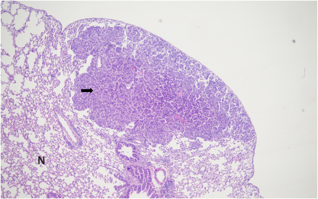
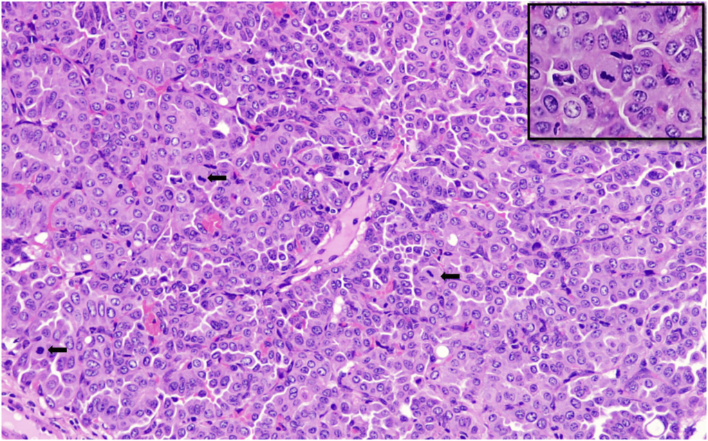
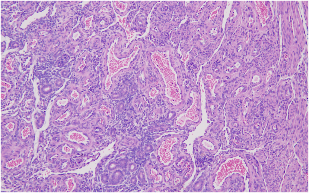
Bronchiolo-alveolar adenomas of the lungs were the most frequently observed spontaneous neoplasms in both male and female mice in the present study, with incidences of 10% in males and 8% in females. These findings are consistent with previously published data by Paranjpe et al,7 which reported incidences of 11.69% and 8.51% in male mice and 6.47% and 6.35% in female mice across two study periods.
In comparison, Labcorp16 reported lower incidences of bronchiolo-alveolar adenomas in males (6.3%) and females (2.3%), suggesting a reduced background occurrence in their data relative to our findings.
In our study, bronchiolo-alveolar adenocarcinoma was observed in 4% of male mice and was absent in female mice. These findings were compared with historical data from Paranjpe et al and Labcorp. Paranjpe et al7 reported lower incidences in male mice of 0.56% and 2.21% and higher incidences in female mice of 1.27% and 2.00% across the two study periods. The data from Labcorp showed an incidence of 1.6% in male mice, which was lower than our findings, and 0.5% in female mice,16 which was consistent with our results.
Overall, the incidence of bronchiolo-alveolar adenocarcinomas was notably lower than that of bronchiolo-alveolar adenomas across all datasets, including the current study.
The combined incidence of bronchiolo-alveolar adenomas and adenocarcinomas in the lungs was 14% in male mice and 8% in female mice in the present study. This finding of total lung tumors is consistent with the data reported by Paranjpe et al,7 which documented combined incidences of 12.25% and 10.72% in male mice and 7.75% and 8.35% in female mice across the two study periods. In contrast, Labcorp reported lower combined incidences of 7.9% in males and 2.8% in females, suggesting a reduced background rate of spontaneous neoplastic findings of lungs in their studies.16
The incidence of bronchiolo-alveolar adenomas of the lungs (10% in males and 8% in females) was also compared with other published data wherein the earlier data from CIEA18 reported slightly lower incidences of 5% in males and 6% in females, while more recent data from CLEA18 showed higher rates of 11.6% in males and 11.1% in females, closely aligning with our observations.
In contrast, bronchiolo-alveolar adenocarcinomas were less frequent. The current study reported an incidence of 4% in males, which was lower than the 7% observed in Central Institute for Experimental Animals (CIEA)17 data and absent in the Central Laboratory for Experimental Animals (CLEA) dataset. Similarly, in females there was no incidence of bronchiolo-alveolar adenocarcinomas in the current study which was in close alignment of 0.6% incidence as observed in the CLEA,18 while the CIEA data reported an incidence of 3% in female mice.
When considering the combined incidence of lung tumors (adenomas and adenocarcinomas), the present study reported 14%, which aligns closely with CIEA at 12% and CLEA at 11.6%. This overall consistency supports the reliability of lung tumor data across all these studies.
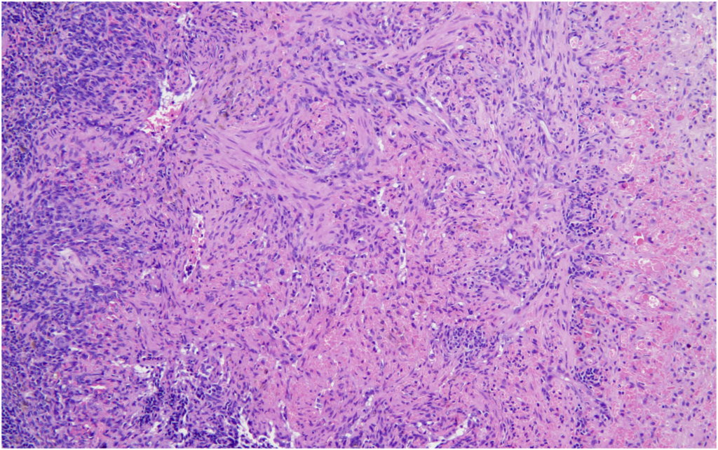
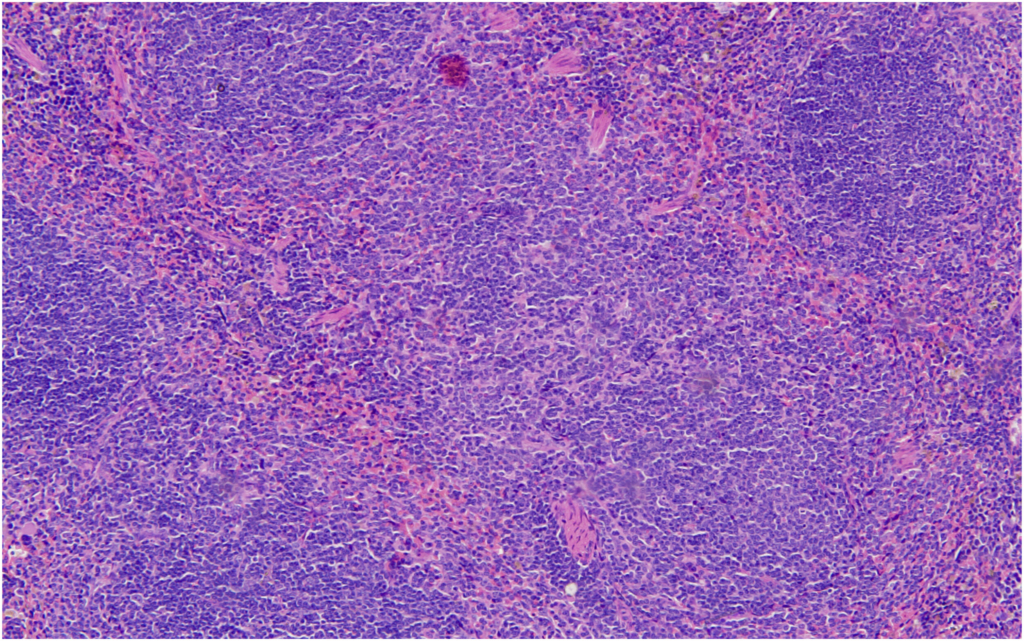
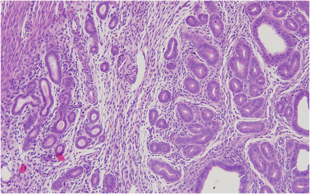
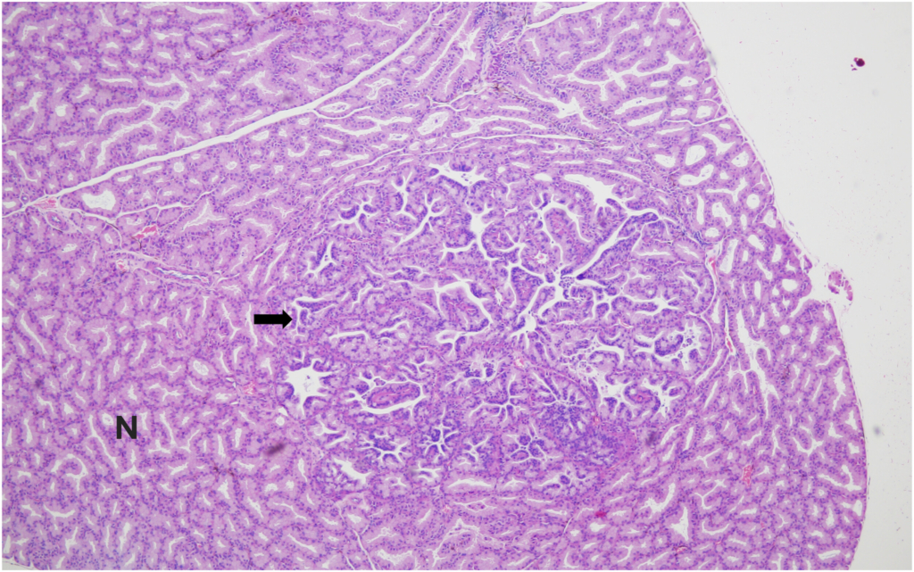
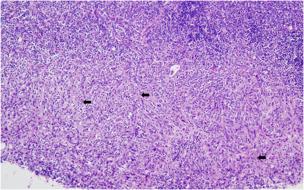
Hemangiosarcoma of the spleen was identified as the second most common spontaneous neoplasm in both male and female mice in the present study. The incidence was 2% in male mice and 4% in female mice, aligning closely with previously reported data. Historical data from Paranjpe et al documented slightly higher incidences of 3.66% and 3.87% in male mice and 3.66% and 3.76% in female mice, respectively. Similarly, Labcorp reported incidences of 3.8% in males and 3.5% in females, further supporting the consistency of this lesion across studies.
The incidence of splenic hemangiosarcoma when compared with other data from mice sourced from Japan revealed that the historical data from CIEA reported higher incidences of 8% in males and 7% in females, while more recent data from CLEA showed an incidence of 4.4% in males and 6% in female mice.
These comparisons suggest that while the incidence of splenic hemangiosarcoma was less in our study (2 % males and 4% in females), it remains a reproducible and prominent background neoplastic lesion in these mice.
It is known that Tg.rasH2 mice also occasionally develop hemangiomas and hemangiosarcomas in organs other than the spleen. These tumors present grossly as blood-filled nodules or masses. The microscopic features of these hemangiosarcomas are very similar to the ones described for the spleen above. No hemangiomas were observed in any of the mice. Hemangiosarcoma was also observed in the testes/uterus, with an incidence of 2% each, while its average incidence in the referred publications was found to be lesser in males (0.7% and 0.22%, respectively) and somewhat variable in females (1.41% and 0.12%, respectively). However, it is worth noting that the two publications of Paranjpe et al also describe the “range” of % incidence of all tumors in the studies they used for creating their historical database, and the % range per study for hemangiosarcoma in testes was reported to be 0%–4% while that in the uterus was 0%–8% and 0%–4%.5 Hemangiosarcoma in the testes was also reported to be 1.1% while that in the uterus it was 2% as reported by the CLEA.18 Hemangiosarcoma in the testes and uterus was not reported by Labcorp.16
Accordingly, the incidence of adenocarcinoma of the Harderian gland in male mice in our study was 2%, while its average incidence was 0.14% and 1.44%, respectively, in the two publications and had a range of 0%–4% and 0%–12%, respectively.5 Similarly, the reported incidence of the adenocarcinoma of the Harderian gland in male mice was 0.3% and 5%, respectively, as reported by Labcorp and CIEA.
Although there was no incidence of adenoma of the Harderian gland in male mice in our study, its average was 1.41% and 1.33%, respectively, in the two publications, with a range of 0%–8%. The adenoma of the Harderian gland in female mice had a 2% incidence in our study, and it was comparable with the 2.81% and 1.29% average incidence of this tumor in the two publications (range, 0%–16% and 0%–8%), respectively.5 The incidence of adenoma of the Harderian gland was 0.7% and 1.4% in male and female mice, respectively, as reported by Labcorp.16
Similarly, the reported incidence of the adenoma of the Harderian gland in female mice was 1% as reported by the CIEA.
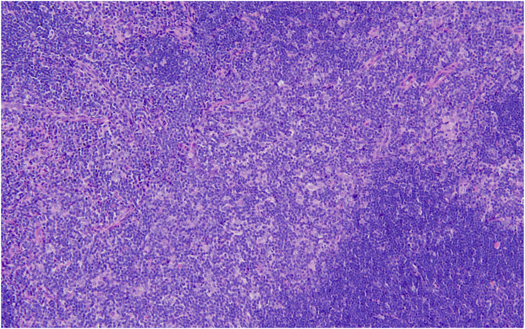
17
Likewise, our study revealed a 2% incidence of thymoma in female mice, as compared to the 0.56% and 1.06% average incidence in the two publications by Paranjpe et al, respectively (range, 0%–12%). Comparative data from Labcorp indicated a thymoma incidence of 0.7% in male mice and 2.2% in female mice, placing our findings in close agreement with their female cohort.
In our study, the presence of squamous cell carcinoma of the salivary glands was noted only in one female mouse (incidence of 2%). The corresponding average incidence of this tumor in female mice, as cited by Paranjpe et al, was 0.14% and 0.35%, respectively (range, 0%–4%). The absence of this tumor in male mice in our study was in agreement with the one of the databases of Paranjpe et al,7 while their other database reported a single male mouse with this tumor (average incidence of 0.14%) when compared across two study periods. Similarly, squamous cell carcinoma of the salivary gland either in male or female mice was not reported in the data published by Labcorp, CLEA, and CIEA.
However, there were a few other tumors that were observed only in the present study conducted at Intox on the mice sourced from CLEA, Japan, and were missing from the two historical control databases from the mice sourced from Taconic, USA, or as reported by other authors in their published data. These included one keratoacanthoma in skin of the head (females) and one rhabdomyoma of the skeletal muscles.
Above discussions reveal that despite the variables such as the wide range of the study conducted dates by Paranjpe et al,7 or by Labcorp,16 geographical locations, number of mice in study, rodent feed, and husbandry practices across the institutions, the tumor spectrum in our study was remarkably similar to these studies. The common spontaneous tumors noted in rasH2 mice sourced from Taconic Inc., USA, were pulmonary adenomas/carcinomas, splenic hemangiosarcomas, and Harderian gland adenomas/carcinomas. Two exceptions noted in our study were the isolated instances of keratoacanthoma of skin and rhabdomyoma in skeletal muscles. The similarity of the incidence of the spontaneous tumors as observed predominantly in the lungs and spleen in this study and when compared with a large database collected over a long-time interval also demonstrate the robustness and minimal drift in this animal model.
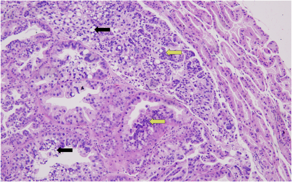
The tumors observed in urethane-treated male and female mice in this study composed of 100% incidence of bronchiolo-alveolar adenomas and 70% incidence of bronchiolo-alveolar adenocarcinomas in lungs (males, 30%; females, 40%), while there was 100% incidence of hemangiosarcomas in the spleen. Similar observations were made in published studies that used urethane as the positive control, which showed a 100% incidence of bronchiolo-alveolar adenomas in Tg.rasH2 mice and adenocarcinomas (males, 30%; females 22.22%) and splenic hemangiomas (20% in males only)/hemangiosarcomas (males, 80%; females 60%). These findings thus validated the use of urethane as a positive control agent in Tg.rasH2 mice, as an alternative to MNU.8

Conclusion

We tested and demonstrated that the incidence of predominant neoplastic findings, both spontaneous and those induced by the positive control urethane, in CB6F1-Tg-rasH2 mice sourced from CLEA Japan Inc, was comparable to that in the mice sourced from Taconic Inc., USA, based on the published databases.